Cardiac Output copertina

Cardiac Output

Cardiac Output

Di: Dr Mike Charlesworth
Ascolta gratuitamente

Offerte di stagione | 0,99 €/mese per i primi 3 mesi

A seguire 9,99 €/mese – si applicano condizioni. Puoi disdire mensilmente.

Cardiac Output is a podcast on cardiothoracic anaesthesia and intensive care medicine.

Dr Mike Charlesworth and Dr Calum Downes bring you tacit knowledge from a national transplant and ECMO centre — the reasoning that never makes it into the textbook.

Copyright 2024 All rights reserved.
  • Cardiac Output Monitoring: Believe the Patient
    Oct 2 2026
    Day one after mitral valve surgery. The monitor says the cardiac index is a healthy three point two. The lactate is six, the feet are cold and the urine has stopped. One of those numbers is lying — and it isn't the lactate. This episode is cardiac output monitoring, for the exam and for the bedside: how each device works, the physiology underneath, when we actually use them, and where each one misleads you. We start with first principles: cardiac output as heart rate times stroke volume, the exam definitions of preload, afterload and contractility, and why a normal blood pressure can hide a failing pump. Then the Fick principle, what a central venous saturation can and can't tell you, and how to diagnose a low-output state by looking at the whole patient rather than one number. Then the devices, one by one. The pulmonary artery catheter — Stewart–Hamilton thermodilution, the continuous thermal-filament version, floating one by the waveforms, complications, the evidence from ESCAPE and PAC-Man, and why it's still the only monitor that shows you the right heart and the pulmonary circulation directly. PiCCO and LiDCO, and their volumetric extras. Uncalibrated pulse contour monitors, and the arrhythmias, valve leaks, vasoplegia and balloon pumps that break them. Stroke volume and pulse pressure variation, and why an open chest and a failing right ventricle make them unreliable. The oesophageal Doppler — the four assumptions behind its number. Echo, which gives you not just the number but the reason. Bioimpedance, and why it never caught on. And what the console really tells you on ECMO, a BiVAD or an implanted LVAD. We finish with a quick-fire quiz, so you can test yourself on the lot. Chapters (00:00) Cold open — one of these numbers is lying(00:50) The physiology: stroke volume, pressure and flow(01:50) The Fick principle and venous saturations(02:55) Diagnosing low output at the bedside(03:55) The pulmonary artery catheter and thermodilution(05:55) Floating a PA catheter by the waveforms(07:25) Complications and the evidence(09:05) Transpulmonary thermodilution — PiCCO(10:20) Lithium dilution — LiDCO(10:55) Uncalibrated pulse contour monitors(12:00) Stroke volume and pulse pressure variation(12:55) The oesophageal Doppler and its assumptions(15:30) Echo: the number and the reason(16:20) Bioimpedance and bioreactance(16:55) Flow on ECMO, BiVADs and LVADs(17:35) Putting it together(18:05) Quick-fire quiz Key takeaways Cardiac output = heart rate × stroke volume; mean arterial pressure = cardiac output × SVR — so a normal pressure says nothing about which one is holding it upCentral venous saturation is not truly mixed venous, and it can mislead in both directions — low for reasons other than output, and high when the tissues can't extract oxygenDiagnose low output from the whole patient: symptoms, signs, end-organ function, lactate and acidosisThe PA catheter is the reference standard (Stewart–Hamilton thermodilution); continuous versions use a thermal filament and lag behind sudden changeKnow the waveforms — RA 0–8, RV 15–30/0–8, PA 15–30/4–12, wedge ~12 mmHg; never leave the balloon wedged; beware existing left bundle branch blockESCAPE and PAC-Man showed no routine survival benefit — use it where the numbers will change what you doPiCCO adds global end-diastolic volume and extravascular lung water, but needs regular recalibration; any thermodilution is unreliable on ECMOUncalibrated pulse contour is good for trends, poor in AF, aortic regurgitation, vasoplegia, a damped line or a balloon pumpSVV/PPV above ~12–13% predicts fluid responsiveness only in a fully ventilated, closed-chest patient in sinus rhythmOesophageal Doppler assumes a nomogram aortic area, a fixed 70:30 split, the right vessel at a sensible angle, and laminar flow; normal FTc 330–360 msEcho stroke volume = LVOT area × VTI — the diameter is squared, so small errors grow; its real value is telling you why the output is lowOn ECMO or a BiVAD the console shows circuit flow only; an LVAD estimates flow from power and speed References / further reading Binanay C et al. Evaluation study of congestive heart failure and pulmonary artery catheterization effectiveness: the ESCAPE trial. JAMA 2005; 294: 1625–33Harvey S et al. Assessment of the clinical effectiveness of pulmonary artery catheters in management of patients in intensive care (PAC-Man): a randomised controlled trial. Lancet 2005; 366: 472–7Marik PE et al. Dynamic changes in arterial waveform derived variables and fluid responsiveness in mechanically ventilated patients: a systematic review of the literature. Crit Care Med 2009; 37: 2642–7Cecconi M et al. Consensus on circulatory shock and hemodynamic monitoring. Task force of the European Society of Intensive Care Medicine. Intensive Care Med 2014; 40: 1795–815Teboul JL et al. Less invasive hemodynamic monitoring in critically ill patients. Intensive Care Med 2016...
    Mostra di più Mostra meno
    19 min
  • Cardioplegia: Stopping the Heart Safely
    Sep 27 2026
    Forty minutes into an aortic valve replacement, the cross-clamp is on and the heart is arrested. Then a little electrical activity starts creeping back on the ECG. What do you do? Tell the surgeon — out loud, straight away — because that heart is waking up while it's still ischaemic. The anaesthetist is part of myocardial protection, even though the solution runs through the perfusionist's pump. This episode is cardioplegia from the top end of the table. We start with why it exists: the moment the aorta is clamped, the heart has no coronary flow, and a warm, beating, ischaemic heart runs out of energy in minutes. Cardioplegia makes planned ischaemia survivable in five ways — arrest, cool, empty, feed and buffer, and limit reperfusion injury — and arrest does most of the work. Then what's in the bag, grouped the way examiners like it: an arresting agent, buffers, osmotic agents and substrate. How potassium produces a depolarised arrest by inactivating fast sodium channels, and why that isn't quite a resting membrane. Why the commonest solution is 4:1 blood cardioplegia, and what blood adds over crystalloid. Warm versus cold as a real trade-off — and the hot shot that gets the heart ready before the clamp comes off. Delivery routes next. Antegrade into the aortic root as standard, direct into the ostia, or down the vein grafts. Retrograde through the coronary sinus for aortic regurgitation, aortic and root surgery, and severe coronary disease — with the 40 mmHg limit on coronary sinus pressure, why retrograde protects the right ventricle less well, and how to confirm the cannula on TOE. And why antegrade into a regurgitant root distends the ventricle instead of perfusing it. Then the seven complications — trauma, air, oedema, bleeding, difficulty restarting, electrolyte disturbance and inadequate protection — and how the last one really shows up: as a heart that comes off badly. del Nido, with its single long-acting dose, lidocaine and low calcium. Custodiol (HTK), which arrests the heart by taking the sodium away rather than adding potassium, and the price you pay in volume and dilution. And Laplace's law, which explains why a distended ventricle is dangerous and why we vent — during surgery, with a balloon pump, and on VA-ECMO. We finish with the anaesthetist's checklist during the cross-clamp, and what to be ready for when it comes off. Chapters (00:00) Cold open — the ECG wakes up during the cross-clamp(01:00) Why we need cardioplegia, and the five mechanisms(01:50) How much oxygen arrest and cooling save(02:30) What's in the bag(02:40) Potassium and the depolarised arrest(04:10) Buffers, osmotic agents and substrate(04:50) Blood versus crystalloid(05:40) Warm versus cold(06:30) The hot shot(07:00) Antegrade delivery(07:40) Retrograde delivery, and when you need it(08:00) Why antegrade fails with aortic regurgitation(08:40) The coronary sinus pressure limit, and the right ventricle(09:20) Confirming the retrograde cannula on TOE(10:00) The seven complications(12:20) del Nido(13:30) Custodiol (HTK)(15:00) Laplace's law, and why we vent(16:00) The anaesthetist's checklist during the cross-clamp(16:30) Wrap-up Key takeaways If electrical activity returns on the ECG during the cross-clamp, tell the surgeon straight away — the heart is working while ischaemicCardioplegia protects the heart in five ways: arrest, cool, empty, deliver substrate and buffering, and limit reperfusion injury — arrest does the heavy liftingContents fall into four groups: an arresting agent (usually potassium, with magnesium), buffers, osmotic agents and substrateHigh potassium raises the resting membrane potential and inactivates fast sodium channels — a depolarised arrest in diastoleBlood cardioplegia (4:1) adds oxygen carriage, buffering, free-radical scavenging and better microvascular flow, with less oedemaCold maximises metabolic suppression; warm supports aerobic metabolism; a warm "hot shot" before clamp removal prepares the heart for reperfusionRetrograde delivery via the coronary sinus is used for significant aortic regurgitation, aortic valve and root surgery, and severe coronary disease — keep sinus pressure below 40 mmHgRetrograde protects the right ventricle less well, so it's often combined with antegradeIn aortic regurgitation, antegrade root delivery distends the left ventricle — watch the LV on TOE and say if it's getting biggerComplications: trauma, air, myocardial oedema, bleeding, difficulty restarting, systemic electrolyte disturbance and inadequate protectiondel Nido (1 part blood to 4 crystalloid, with lidocaine and low calcium) gives a single dose lasting up to around 90 minutesCustodiol (HTK) arrests by sodium depletion — a hyperpolarised arrest — with long protection, but large volumes cause haemodilution and hyponatraemiaLaplace: wall tension rises with pressure and radius — which is why distension is dangerous and why we ventSolutions, doses and protocols are ...
    Mostra di più Mostra meno
    19 min
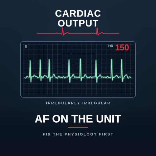
  • AF on the Unit: Fix the Physiology First
    Sep 25 2026
    Eight o'clock ward round, day two after a coronary artery bypass, and the monitor shows atrial fibrillation at a hundred and fifty. Calum's answer is amiodarone — and that is the single most common wrong answer on the unit. Not because amiodarone is wrong, but because of where it sits in the order. This is atrial fibrillation on the cardiothoracic intensive care unit, top to bottom. And it matters beyond the bedside: in a recent Fellowship paper it was the worst-performing question on the whole exam, with a pass rate of about 43 per cent. The examiners were specific about where candidates fell down — the management, and the anaesthetic for elective DC cardioversion. The bit everyone assumes is easy. We start with how common it is — about a third of patients after coronary surgery, up to half after valve or combined procedures, peaking on days two and three — and why it isn't benign. Then a framework you can use on a ward round: substrate and trigger. The substrate is atrial; the triggers are catecholamines, pain, hypovolaemia, hypoxia, anaemia, electrolytes and the inflammatory response to bypass. Almost every trigger is something you can change. Then the four strands of management — rhythm, rate, recurrence and thromboembolism — and the step that comes before all of them. Correct the physiology first: potassium, magnesium, pain, volume; exclude tamponade and pneumothorax with a scan and a film, not a glance at the drain; look for sepsis; and check for a misplaced epicardial pacing wire irritating the atrium. Only then a drug. Amiodarone as the workhorse — why it acts on both rhythm and rate, and why the slow loading dose is a safety feature rather than fussiness in a shocked post-cardiotomy patient. Then the drugs we avoid, and the specific reason for each: digoxin, verapamil and diltiazem, flecainide and the lesson of the CAST trial, beta-blockers (and why esmolol if you must), and adenosine as a diagnostic rather than a treatment. Plus the Vaughan Williams classification in ninety seconds, and when an unstable patient needs a synchronised shock rather than a drug. Anticoagulation next: our local position on new-onset post-operative AF and the reasoning behind it, anticoagulant mechanisms and reversal agents, the case for and against the direct oral anticoagulants, and neuraxial timings. Then a trap specific to cardiac surgery — andexanet alfa can look exactly like heparin resistance on bypass, which is why you tell the perfusionist before you reverse, not afterwards. We finish with the section the examiners said was done worst — the anaesthetic for a DC cardioversion list, including what to avoid and the bailout you set up before the first patient — and the long-term options for rhythm, rate and stroke prevention. Chapters (00:00) Cold open — AF at 150, and the commonest wrong answer(00:50) Why this was the worst-answered exam question(01:20) Incidence, and why it isn't benign(02:10) Substrate and trigger(02:40) Causes of AF in general(03:30) The four strands of management(03:50) Correct the physiology first(04:20) The misplaced pacing wire(04:50) Amiodarone, and why the slow load matters(06:30) The drugs we avoid — and why(08:00) The Vaughan Williams classification(08:50) The unstable patient(09:20) Anticoagulation: our local position(10:00) Mechanisms and reversal(11:00) For and against the DOACs(11:30) Neuraxial timings(11:50) Reversal before bypass — andexanet and apparent heparin resistance(13:10) Anaesthesia for DC cardioversion(14:30) Long-term rhythm, rate and stroke prevention(15:10) Wrap-up Key takeaways Post-operative AF affects about a third of patients after CABG and up to half after valve or combined surgery, peaking on days two to three — often self-terminating, but not benignThink substrate (atrial trauma, inflammation, stretch, fibrosis, a large left atrium) and trigger (catecholamines, pain, hypovolaemia, hypoxia, anaemia, electrolytes, the inflammatory response)Four strands of management: restore rhythm, control rate, prevent recurrence, prevent thromboembolismCorrect the physiology first: K⁺ 4.5–5.5, Mg²⁺ above 1.0, treat pain and hypovolaemia, exclude tamponade and pneumothorax, look for sepsis, and check the pacing wiresAmiodarone 300 mg over 20–60 minutes (never as a rapid bolus), centrally where possible, then 600–900 mg over 24 hours — rapid loading causes negative inotropy and vasodilationDigoxin is not an acute agent; verapamil and diltiazem are negatively inotropic; flecainide is contraindicated in structural heart disease and ischaemia (the CAST lesson); esmolol if you must beta-block; adenosine is diagnostic, not therapeuticAdverse features — shock, syncope, myocardial ischaemia, heart failure — mean synchronised DC cardioversion, not a drug; know the Resuscitation Council adult tachycardia algorithmKnow anticoagulants by mechanism, because reversal follows mechanismReversing a DOAC before bypass? Tell the surgeon and the ...
    Mostra di più Mostra meno
    20 min
adbl_web_anon_alc_button_suppression_t1
Ancora nessuna recensione