Episodi

  • Cardiac Output Monitoring: Believe the Patient
    Oct 2 2026
    Day one after mitral valve surgery. The monitor says the cardiac index is a healthy three point two. The lactate is six, the feet are cold and the urine has stopped. One of those numbers is lying — and it isn't the lactate. This episode is cardiac output monitoring, for the exam and for the bedside: how each device works, the physiology underneath, when we actually use them, and where each one misleads you. We start with first principles: cardiac output as heart rate times stroke volume, the exam definitions of preload, afterload and contractility, and why a normal blood pressure can hide a failing pump. Then the Fick principle, what a central venous saturation can and can't tell you, and how to diagnose a low-output state by looking at the whole patient rather than one number. Then the devices, one by one. The pulmonary artery catheter — Stewart–Hamilton thermodilution, the continuous thermal-filament version, floating one by the waveforms, complications, the evidence from ESCAPE and PAC-Man, and why it's still the only monitor that shows you the right heart and the pulmonary circulation directly. PiCCO and LiDCO, and their volumetric extras. Uncalibrated pulse contour monitors, and the arrhythmias, valve leaks, vasoplegia and balloon pumps that break them. Stroke volume and pulse pressure variation, and why an open chest and a failing right ventricle make them unreliable. The oesophageal Doppler — the four assumptions behind its number. Echo, which gives you not just the number but the reason. Bioimpedance, and why it never caught on. And what the console really tells you on ECMO, a BiVAD or an implanted LVAD. We finish with a quick-fire quiz, so you can test yourself on the lot. Chapters (00:00) Cold open — one of these numbers is lying(00:50) The physiology: stroke volume, pressure and flow(01:50) The Fick principle and venous saturations(02:55) Diagnosing low output at the bedside(03:55) The pulmonary artery catheter and thermodilution(05:55) Floating a PA catheter by the waveforms(07:25) Complications and the evidence(09:05) Transpulmonary thermodilution — PiCCO(10:20) Lithium dilution — LiDCO(10:55) Uncalibrated pulse contour monitors(12:00) Stroke volume and pulse pressure variation(12:55) The oesophageal Doppler and its assumptions(15:30) Echo: the number and the reason(16:20) Bioimpedance and bioreactance(16:55) Flow on ECMO, BiVADs and LVADs(17:35) Putting it together(18:05) Quick-fire quiz Key takeaways Cardiac output = heart rate × stroke volume; mean arterial pressure = cardiac output × SVR — so a normal pressure says nothing about which one is holding it upCentral venous saturation is not truly mixed venous, and it can mislead in both directions — low for reasons other than output, and high when the tissues can't extract oxygenDiagnose low output from the whole patient: symptoms, signs, end-organ function, lactate and acidosisThe PA catheter is the reference standard (Stewart–Hamilton thermodilution); continuous versions use a thermal filament and lag behind sudden changeKnow the waveforms — RA 0–8, RV 15–30/0–8, PA 15–30/4–12, wedge ~12 mmHg; never leave the balloon wedged; beware existing left bundle branch blockESCAPE and PAC-Man showed no routine survival benefit — use it where the numbers will change what you doPiCCO adds global end-diastolic volume and extravascular lung water, but needs regular recalibration; any thermodilution is unreliable on ECMOUncalibrated pulse contour is good for trends, poor in AF, aortic regurgitation, vasoplegia, a damped line or a balloon pumpSVV/PPV above ~12–13% predicts fluid responsiveness only in a fully ventilated, closed-chest patient in sinus rhythmOesophageal Doppler assumes a nomogram aortic area, a fixed 70:30 split, the right vessel at a sensible angle, and laminar flow; normal FTc 330–360 msEcho stroke volume = LVOT area × VTI — the diameter is squared, so small errors grow; its real value is telling you why the output is lowOn ECMO or a BiVAD the console shows circuit flow only; an LVAD estimates flow from power and speed References / further reading Binanay C et al. Evaluation study of congestive heart failure and pulmonary artery catheterization effectiveness: the ESCAPE trial. JAMA 2005; 294: 1625–33Harvey S et al. Assessment of the clinical effectiveness of pulmonary artery catheters in management of patients in intensive care (PAC-Man): a randomised controlled trial. Lancet 2005; 366: 472–7Marik PE et al. Dynamic changes in arterial waveform derived variables and fluid responsiveness in mechanically ventilated patients: a systematic review of the literature. Crit Care Med 2009; 37: 2642–7Cecconi M et al. Consensus on circulatory shock and hemodynamic monitoring. Task force of the European Society of Intensive Care Medicine. Intensive Care Med 2014; 40: 1795–815Teboul JL et al. Less invasive hemodynamic monitoring in critically ill patients. Intensive Care Med 2016...
    Mostra di più Mostra meno
    19 min
  • Cardioplegia: Stopping the Heart Safely
    Sep 27 2026
    Forty minutes into an aortic valve replacement, the cross-clamp is on and the heart is arrested. Then a little electrical activity starts creeping back on the ECG. What do you do? Tell the surgeon — out loud, straight away — because that heart is waking up while it's still ischaemic. The anaesthetist is part of myocardial protection, even though the solution runs through the perfusionist's pump. This episode is cardioplegia from the top end of the table. We start with why it exists: the moment the aorta is clamped, the heart has no coronary flow, and a warm, beating, ischaemic heart runs out of energy in minutes. Cardioplegia makes planned ischaemia survivable in five ways — arrest, cool, empty, feed and buffer, and limit reperfusion injury — and arrest does most of the work. Then what's in the bag, grouped the way examiners like it: an arresting agent, buffers, osmotic agents and substrate. How potassium produces a depolarised arrest by inactivating fast sodium channels, and why that isn't quite a resting membrane. Why the commonest solution is 4:1 blood cardioplegia, and what blood adds over crystalloid. Warm versus cold as a real trade-off — and the hot shot that gets the heart ready before the clamp comes off. Delivery routes next. Antegrade into the aortic root as standard, direct into the ostia, or down the vein grafts. Retrograde through the coronary sinus for aortic regurgitation, aortic and root surgery, and severe coronary disease — with the 40 mmHg limit on coronary sinus pressure, why retrograde protects the right ventricle less well, and how to confirm the cannula on TOE. And why antegrade into a regurgitant root distends the ventricle instead of perfusing it. Then the seven complications — trauma, air, oedema, bleeding, difficulty restarting, electrolyte disturbance and inadequate protection — and how the last one really shows up: as a heart that comes off badly. del Nido, with its single long-acting dose, lidocaine and low calcium. Custodiol (HTK), which arrests the heart by taking the sodium away rather than adding potassium, and the price you pay in volume and dilution. And Laplace's law, which explains why a distended ventricle is dangerous and why we vent — during surgery, with a balloon pump, and on VA-ECMO. We finish with the anaesthetist's checklist during the cross-clamp, and what to be ready for when it comes off. Chapters (00:00) Cold open — the ECG wakes up during the cross-clamp(01:00) Why we need cardioplegia, and the five mechanisms(01:50) How much oxygen arrest and cooling save(02:30) What's in the bag(02:40) Potassium and the depolarised arrest(04:10) Buffers, osmotic agents and substrate(04:50) Blood versus crystalloid(05:40) Warm versus cold(06:30) The hot shot(07:00) Antegrade delivery(07:40) Retrograde delivery, and when you need it(08:00) Why antegrade fails with aortic regurgitation(08:40) The coronary sinus pressure limit, and the right ventricle(09:20) Confirming the retrograde cannula on TOE(10:00) The seven complications(12:20) del Nido(13:30) Custodiol (HTK)(15:00) Laplace's law, and why we vent(16:00) The anaesthetist's checklist during the cross-clamp(16:30) Wrap-up Key takeaways If electrical activity returns on the ECG during the cross-clamp, tell the surgeon straight away — the heart is working while ischaemicCardioplegia protects the heart in five ways: arrest, cool, empty, deliver substrate and buffering, and limit reperfusion injury — arrest does the heavy liftingContents fall into four groups: an arresting agent (usually potassium, with magnesium), buffers, osmotic agents and substrateHigh potassium raises the resting membrane potential and inactivates fast sodium channels — a depolarised arrest in diastoleBlood cardioplegia (4:1) adds oxygen carriage, buffering, free-radical scavenging and better microvascular flow, with less oedemaCold maximises metabolic suppression; warm supports aerobic metabolism; a warm "hot shot" before clamp removal prepares the heart for reperfusionRetrograde delivery via the coronary sinus is used for significant aortic regurgitation, aortic valve and root surgery, and severe coronary disease — keep sinus pressure below 40 mmHgRetrograde protects the right ventricle less well, so it's often combined with antegradeIn aortic regurgitation, antegrade root delivery distends the left ventricle — watch the LV on TOE and say if it's getting biggerComplications: trauma, air, myocardial oedema, bleeding, difficulty restarting, systemic electrolyte disturbance and inadequate protectiondel Nido (1 part blood to 4 crystalloid, with lidocaine and low calcium) gives a single dose lasting up to around 90 minutesCustodiol (HTK) arrests by sodium depletion — a hyperpolarised arrest — with long protection, but large volumes cause haemodilution and hyponatraemiaLaplace: wall tension rises with pressure and radius — which is why distension is dangerous and why we ventSolutions, doses and protocols are ...
    Mostra di più Mostra meno
    19 min
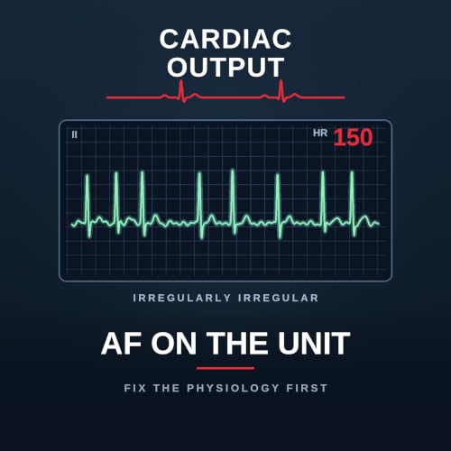
  • AF on the Unit: Fix the Physiology First
    Sep 25 2026
    Eight o'clock ward round, day two after a coronary artery bypass, and the monitor shows atrial fibrillation at a hundred and fifty. Calum's answer is amiodarone — and that is the single most common wrong answer on the unit. Not because amiodarone is wrong, but because of where it sits in the order. This is atrial fibrillation on the cardiothoracic intensive care unit, top to bottom. And it matters beyond the bedside: in a recent Fellowship paper it was the worst-performing question on the whole exam, with a pass rate of about 43 per cent. The examiners were specific about where candidates fell down — the management, and the anaesthetic for elective DC cardioversion. The bit everyone assumes is easy. We start with how common it is — about a third of patients after coronary surgery, up to half after valve or combined procedures, peaking on days two and three — and why it isn't benign. Then a framework you can use on a ward round: substrate and trigger. The substrate is atrial; the triggers are catecholamines, pain, hypovolaemia, hypoxia, anaemia, electrolytes and the inflammatory response to bypass. Almost every trigger is something you can change. Then the four strands of management — rhythm, rate, recurrence and thromboembolism — and the step that comes before all of them. Correct the physiology first: potassium, magnesium, pain, volume; exclude tamponade and pneumothorax with a scan and a film, not a glance at the drain; look for sepsis; and check for a misplaced epicardial pacing wire irritating the atrium. Only then a drug. Amiodarone as the workhorse — why it acts on both rhythm and rate, and why the slow loading dose is a safety feature rather than fussiness in a shocked post-cardiotomy patient. Then the drugs we avoid, and the specific reason for each: digoxin, verapamil and diltiazem, flecainide and the lesson of the CAST trial, beta-blockers (and why esmolol if you must), and adenosine as a diagnostic rather than a treatment. Plus the Vaughan Williams classification in ninety seconds, and when an unstable patient needs a synchronised shock rather than a drug. Anticoagulation next: our local position on new-onset post-operative AF and the reasoning behind it, anticoagulant mechanisms and reversal agents, the case for and against the direct oral anticoagulants, and neuraxial timings. Then a trap specific to cardiac surgery — andexanet alfa can look exactly like heparin resistance on bypass, which is why you tell the perfusionist before you reverse, not afterwards. We finish with the section the examiners said was done worst — the anaesthetic for a DC cardioversion list, including what to avoid and the bailout you set up before the first patient — and the long-term options for rhythm, rate and stroke prevention. Chapters (00:00) Cold open — AF at 150, and the commonest wrong answer(00:50) Why this was the worst-answered exam question(01:20) Incidence, and why it isn't benign(02:10) Substrate and trigger(02:40) Causes of AF in general(03:30) The four strands of management(03:50) Correct the physiology first(04:20) The misplaced pacing wire(04:50) Amiodarone, and why the slow load matters(06:30) The drugs we avoid — and why(08:00) The Vaughan Williams classification(08:50) The unstable patient(09:20) Anticoagulation: our local position(10:00) Mechanisms and reversal(11:00) For and against the DOACs(11:30) Neuraxial timings(11:50) Reversal before bypass — andexanet and apparent heparin resistance(13:10) Anaesthesia for DC cardioversion(14:30) Long-term rhythm, rate and stroke prevention(15:10) Wrap-up Key takeaways Post-operative AF affects about a third of patients after CABG and up to half after valve or combined surgery, peaking on days two to three — often self-terminating, but not benignThink substrate (atrial trauma, inflammation, stretch, fibrosis, a large left atrium) and trigger (catecholamines, pain, hypovolaemia, hypoxia, anaemia, electrolytes, the inflammatory response)Four strands of management: restore rhythm, control rate, prevent recurrence, prevent thromboembolismCorrect the physiology first: K⁺ 4.5–5.5, Mg²⁺ above 1.0, treat pain and hypovolaemia, exclude tamponade and pneumothorax, look for sepsis, and check the pacing wiresAmiodarone 300 mg over 20–60 minutes (never as a rapid bolus), centrally where possible, then 600–900 mg over 24 hours — rapid loading causes negative inotropy and vasodilationDigoxin is not an acute agent; verapamil and diltiazem are negatively inotropic; flecainide is contraindicated in structural heart disease and ischaemia (the CAST lesson); esmolol if you must beta-block; adenosine is diagnostic, not therapeuticAdverse features — shock, syncope, myocardial ischaemia, heart failure — mean synchronised DC cardioversion, not a drug; know the Resuscitation Council adult tachycardia algorithmKnow anticoagulants by mechanism, because reversal follows mechanismReversing a DOAC before bypass? Tell the surgeon and the ...
    Mostra di più Mostra meno
    20 min
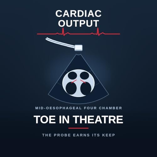
  • TOE in Theatre: The Probe Earns Its Keep
    Sep 21 2026
    Last episode we put a probe on the chest. This one goes down the oesophagus — and it starts somewhere uncomfortable. Inserting a transoesophageal probe is something we do to a patient that can cause them significant harm. Maybe once a year, sometimes more, a patient comes to harm because an anaesthetist put a probe down. We do a great many of these, so it is not unreasonable to think we sometimes cause damage and never find out. But sometimes the injury is severe: an oesophageal tear, upper gastrointestinal bleeding. Severe harm runs at roughly one in two thousand, from a UK audit by the Association of Cardiothoracic Anaesthetists, with female sex, steroid use and a low body mass index as risk factors — all observational, so hold it loosely. And then the argument that follows from it. Contraindications are genuinely hard to find, and Mike's position has changed over time: transoesophageal echo should be mandatory for essentially all cardiac surgery unless there is a good reason not to. Not because every operation demands it, but because of what turns up — valvular problems and dissections in patients listed for bypass grafts with supposedly normal hearts, things that would never have been found without scanning them on the table. From there, the practical half. Three ways to get an ejection fraction, in ascending order of rigour: the eyeball, which in experienced hands correlates well and is what you'll actually use in a crashing patient; fractional area change from the transgastric short axis; and Simpson's biplane, the most rigorous and the most dependent on an endocardial border you can genuinely see. Garbage in, garbage out. Then the right ventricle, and an argument worth having: there are many accepted ways to assess it, and the very existence of so many tells you that none is good enough alone. Watch TAPSE being measured in theatre and ask honestly whether that is a precise, scientific number — particularly off-axis on a transoesophageal probe. Most of the time what you are doing is judging performance by eye and following the trend: how it looked before, how it looks now, what you did in between, and whether it worked. Plus the levers when the right ventricle is struggling — rate and rhythm, preload, afterload, contractility and coronary perfusion, and what comes after them. Systolic anterior motion gets done properly this time, and the key idea is that echo lets you predict it rather than just diagnose it: a long anterior mitral leaflet, a small hypertrophied ventricle with a narrow outflow tract, and a short coaptation-to-septum distance. Then the management, which runs almost exactly opposite to instinct. Then the probe as a theatre instrument. Siting a balloon pump tip just distal to the subclavian — and what goes wrong if it sits too high or too low. The Protek Duo, and why its tip must sit beyond the pulmonary valve. And the rule worth carrying out of the whole topic: any patient on mechanical support with haemodynamic instability does not have to do very much to earn a scan. The section Mike calls most underrated is the one where you help the surgeon and the perfusionist. The venous drainage cannula that has slipped into a hepatic vein — presenting as low flows and poor drainage that everyone assumes is volume — and the non-standard view that finds it. Confirming the retrograde cardioplegia cannula in the coronary sinus, which protects the heart for the entire operation. And watching the wires: the femoral venous wire in peripheral bypass, and the balloon pump wire in the descending aorta, with a live commentary nobody else in the room can give. We finish with transplantation — what is and isn't useful to scan, why a heart that has just been ischaemic cannot be judged like a normal one, and the three questions all of this monitoring exists to answer before you take that patient out to the unit. Chapters (00:00) Cold open — the probe can hurt people(01:00) One in two thousand, and the risk factors(01:40) Contraindications, and how few there are(02:20) Who should get one — and why the answer changed(03:20) Accreditation for cardiac anaesthetists(03:50) Ejection fraction: eyeball, FAC and Simpson's biplane(05:00) The right ventricle, and why so many methods is a warning(06:20) The levers when the right ventricle is failing(07:00) Predicting systolic anterior motion before it happens(08:00) Treating it when it happens(08:40) Siting a balloon pump(09:20) The Protek Duo(10:10) Instability on support earns a scan(11:00) Which pipes you can actually see(11:50) Helping the surgeon: the cannula in the hepatic vein(12:50) Confirming retrograde cardioplegia in the coronary sinus(13:20) Watching the wires(13:50) Transplantation(15:00) Wrap-up Key takeaways Putting a probe down can seriously harm a patient: severe harm is around 1 in 2000, with female sex, steroids and low BMI as observational risk factorsContraindications are few — oesophageal pathology or swallowing difficulty...
    Mostra di più Mostra meno
    15 min
  • Transthoracic Echo on the Unit: Five Views and Six Patterns
    Sep 20 2026
    Three in the morning. A patient on the unit is hypotensive, the noradrenaline is going up, and somebody has already given a litre. What is the fastest way to actually know what's wrong? Put a probe on them. This is the first of two foundation episodes on echocardiography — the thing that turns a guess into a diagnosis, at the bedside, in about ninety seconds. Today it's transthoracic echo on the intensive care unit: the views, and the patterns you are looking for in a shocked patient. Next time we take the transoesophageal probe into theatre. We start with accreditation, because Mike's advice is to start early. The routes come in two shapes and it helps to see that first: a mentored portfolio, or an examination. FICE is the portfolio route — an approved basic course, a logbook of fifty studies with at least ten directly supervised, all fifty reviewed with your mentor, and a triggered assessment, with the first and last scans no more than twelve months apart. EDEC is the examination route — a hundred transthoracic and thirty-five transoesophageal cases, a mentor and a supervisor, and a formal exam you can start once you've done thirty and ten. Plus the machine you actually need: a cardiac phased-array probe, 2D and M-mode, colour and spectral Doppler, ECG gating — and proper image archiving, for three reasons most people haven't thought about. Then the five views that will get you a very long way. Parasternal long axis for overall size and function and the mitral and aortic valves. Parasternal short axis — the doughnut — which at papillary muscle level is the single best view for regional wall motion and for the shape of the septum. Apical four-chamber for comparing the ventricles, for TAPSE, and for Doppler through the mitral and tricuspid valves. Subcostal, the one people neglect and the one that works when nothing else does, because it doesn't care about ventilation, chest drains or dressings — and the best view for a pericardial effusion. And the inferior vena cava from there. Then six patterns. Hypovolaemia, with its hyperdynamic ventricle, end-systolic effacement and kissing walls. Low afterload, which can look almost identical — and why that means echo is never read in isolation. Right ventricular failure, usually obvious the moment the probe goes on: a dilated right ventricle squashing a small, underfilled left one. Tamponade, with diastolic collapse of the right atrium and ventricle. Dynamic left ventricular outflow tract obstruction and systolic anterior motion — which looks like a mitral valve problem and is really a haemodynamic one, where the management runs almost exactly opposite to your instincts. And type A dissection: a normal-looking heart with a flap in the root. The case we'd want every new starter to know is the aortic valve replacement done for aortic stenosis. That hypertrophied ventricle now ejects vigorously through a brand-new valve, empties, and becomes profoundly hypovolaemic. The vasopressor requirement climbs, somebody quite reasonably asks for an echo to exclude tamponade — and the scan shows hypovolaemia instead. That is echo earning its keep: not confirming what you suspected, but telling you that you were looking at the wrong thing. We finish on fluid responsiveness, with some honest context about why it matters less than it used to, the real pitfalls of the vena cava, and why lung ultrasound and venous congestion assessment tell you more than any single number. Chapters (00:00) Cold open — 3am, hypotensive, a litre already in(00:40) Why accreditation is worth starting now(01:30) Two shapes of pathway: FICE and EDEC(03:00) The machine you actually need — and why archiving matters(04:00) The five views: parasternal long axis(04:40) Parasternal short axis — the doughnut(05:30) Apical four-chamber(06:00) Subcostal, and the inferior vena cava(06:50) Hypovolaemia: kissing walls and a collapsed cava(07:40) Why low afterload looks the same(08:20) Right ventricular failure(09:00) Tamponade(09:40) Dynamic LVOT obstruction and systolic anterior motion(10:30) Type A dissection(11:00) The classic case: the scan you ordered for tamponade(12:00) Fluid responsiveness, honestly(13:00) Wrap-up Key takeaways Echo turns a guess into a diagnosis at the bedside in about ninety seconds — put the probe on earlyStart accreditation now: FICE is a mentored portfolio route, EDEC is examination-based, and both take time. Requirements change, so check the current versionYour machine needs a cardiac phased-array probe, 2D and M-mode, colour and spectral Doppler, ECG gating — and image archiving, without which you have no logbook, no comparison and no recordFive views will take you a long way: parasternal long axis, parasternal short axis, apical four-chamber, subcostal, and the inferior vena cavaThe parasternal short axis at papillary muscle level is the best single view for regional wall motion and for septal shapeThe subcostal view is the rescue view — it works ...
    Mostra di più Mostra meno
    14 min
  • Journal Club: GLP-1 Agonists and Aspiration (GLIMPSE)
    Sep 17 2026
    Recorded minutes after the live launch at Annual Congress 2026, and published the same day the paper appears in Anaesthesia. GLIMPSE — the GLP-1 receptor agonist Management in the Peri-operative Setting project — is a national, prospective, multicentre cohort study delivered through the UK resident research networks across all four nations, and it is the largest thing anyone has done on this question. Mike and Calum take it apart the way they did ITACS: the question, the methods, the results, the critique. Then the half that wasn't in the room — what it means for cardiothoracic anaesthesia and intensive care. Two numbers to start. One in 36 patients presenting for anaesthesia in the UK is taking a GLP-1 receptor agonist. And in those patients, the incidence of pulmonary aspiration or regurgitation was 1 in 71, against 1 in 802 in everyone else — an odds ratio of about 11. Then the work of deciding how much of that to believe. The framing that governs everything: the primary outcome is a prevalence, not a comparison. GLIMPSE was designed to answer "how many?" The aspiration finding is a secondary outcome, and every conclusion about the odds ratio has to sit inside that. We cover the design — each site choosing its own fortnight, screening every patient's drug history — the exclusions, the deliberate discarding of twelve sites that screened fewer than three quarters of their eligible patients, and the 97% screening rate that makes this a remarkably complete national snapshot. The prevalence findings are more interesting than the headline. Between institutions, use ranged from 0.4% to 7.8% — a twenty-fold difference — so the national average is close to useless for planning your own list. Tirzepatide accounts for 70% and semaglutide 26%, and this is now predominantly a weight-loss population rather than a diabetes one. And the number that should change your practice this week: 41% obtained the drug from somewhere that is neither a GP nor a hospital specialist. It will not be on the GP summary, the discharge letter or the drug chart. It is only there if somebody asks the patient directly, by name. Management was highly variable. Thirty per cent were asked to stop, most for 8–14 days — consistent with "stop it for a week", which the paper quietly demolishes: with half-lives of five and seven days, five half-lives means 25 and 35 days. A week does not eliminate the drug, and national consensus guidance says to continue it anyway. Meanwhile gastric ultrasound — the one bedside test that answers precisely the question being asked — was used in 1.3% of patients. The safety finding gets the full appraisal, including the qualifier that changes how you read it: of the 19 events in the exposed group, all 19 involved regurgitation and only two reached the lung, which suggests the intubation, head-up positioning and pre-oxygenation were doing their job. And the finding we'd most like to change practice: more than half the events happened at emergence, not induction. The critique covers association without any ability to adjust for confounding, observer bias inflating the ratio from both ends (with the NAP7 cross-check that partly defends it), small event numbers driving a large effect, and generalisability. The strengths get their due too. Then the cardiothoracic half, which the paper does not address. Why our prevalence is probably higher than the national figure. Why the months between assessment and surgery make the drug-history gap worse for us. Why emergence happening on the intensive care unit, hours later, makes this a handover problem as much as an airway problem.What it means that we put a probe into the stomach of nearly every patient we anaesthetise — and that we already own the skill that was used in 1% of cases nationally. The double-lumen tube versus tube-exchange dilemma, taken from a real case in the paper. The conflict between a rapid sequence induction and haemodynamic stability in critical aortic stenosis. Why stopping the drug costs our patients more, through hyperglycaemia and sternal wound infection. And the exclusion that matters most to us: GLIMPSE excluded patients whose airway management began elsewhere — ICU transfers explicitly — so our sickest patients are entirely outside the dataset. Chapters (00:00) Cold open — outside Hall 1A, minutes after the launch(00:50) The two headline numbers(01:30) Why the question needed asking(02:40) Methods: design, sites and the fortnight(03:30) The exclusions — including ICU transfers(04:10) The primary outcome is a prevalence, not a comparison(05:00) Powering, and discarding twelve sites on purpose(05:50) One in 36 — and the twenty-fold variation between hospitals(07:00) Which drugs, and why people were taking them(07:40) Where they got them: the 41% that isn't on any record(08:40) What clinicians did: stopping for a week(09:40) Why a week is pharmacologically meaningless(10:30) Airway management — and gastric ...
    Mostra di più Mostra meno
    22 min
  • TOE for Mechanical Support 2: VADs and the Impella
    Sep 13 2026
    Before we touch a single image, we sort out the vocabulary — because almost everyone muddles these devices, and once you have muddled them you cannot reason about them. People say "VAD" and mean five different machines, sitting in different places, for different lengths of time. So: three questions, and every device answers all three. Which ventricle? How long is it meant to stay in? Surgical or percutaneous? This is part two of two, following the structure of the mechanical support chapter from the fourth edition of Practical Perioperative Transoesophageal Echocardiography. Part one covered balloon pumps and ECMO. Every assist device has the same four components — a pump providing continuous flow, an inflow cannula, an outflow cannula, and an external controller connected by a driveline — and all of them sit in parallel with the native circulation. An LVAD drains from the left ventricular apex and returns to the ascending aorta end to side; an RVAD drains from the right atrium and returns to the pulmonary artery. Temporary means days to weeks, durable means weeks to years, and those are genuinely different machines rather than the same one left in longer. The predominant durable device worldwide is now the HeartMate 3. Then the word that causes the most trouble: BiVAD. Two quite different situations get called the same thing. A true biventricular assist device means two devices implanted for long-term biventricular support, and that is occasional. Whereas a patient receiving a durable LVAD not infrequently needs a temporary RVAD at the same operation, because the right heart cannot cope — two devices, but one durable and one coming out. BiVAD describes the anatomy and tells you nothing about the timescale, which is precisely why people get confused. With that grid in place, the rest follows the chapter. Before implantation: ventricular assessment, intracardiac thrombus, the valves, the aorta, and intracardiac shunts. Shunts matter because of a pressure change you are about to create — once support starts, left atrial pressure falls below right atrial pressure, so a patent foramen that has been silent for a lifetime can shunt right to left, giving arterial hypoxaemia or systemic embolisation of right-sided gas or thrombus. And because a foramen can be genuinely hard to detect before support starts, you re-examine the atrial septum afterwards. Aortic regurgitation gets the attention it deserves, because it is the lesion that quietly destroys LVAD efficacy: blood leaves the outflow cannula, flows back across the incompetent valve, and returns straight into the inflow cannula — a circuit inside the chest that never reaches the patient. How you correct it depends on the intention of the device, why a bioprosthesis is preferred if replacement is needed, why colour Doppler underestimates it in end-stage failure and you should therefore assess on bypass, which measures work and which do not — and a free clue that costs nothing, in the left ventricular vent flows during implantation. Plus the exception on aortic stenosis that matters only for partial-support devices, and the mitral and tricuspid lesions corrected at the same operation. After implantation: de-airing, and then the single most useful structure on the screen. The interventricular septum should be flat and neutral. Marked rightward displacement means the ventricle is inadequately decompressed — underpumping. Marked leftward means it has collapsed — the suction event, or ventricular suckdown. That is how you run a ramp study, and it is the same logic you will use later for the Impella. Right ventricular function afterwards gets four reasons why it does not always improve despite reduced afterload, the echo signs of acute right ventricular failure, and then — in its proper place — temporary right ventricular support: either a surgically grafted CentriMag, or the Protek Duo, a percutaneous dual-lumen cannula from the right internal jugular with its inflow in the right atrium and its outflow in the proximal main pulmonary artery. We then cover aortic valve opening and the HeartMate 3's programmed rhythmic flow changes, cannula assessment including the biplane tip for confirming inflow orientation, early and late causes of obstruction, why you reinterrogate both cannulas at chest closure, how to read the echo alongside the console, and the surveillance schedule. The episode closes with temporary ventricular assist devices as their own topic — the Impella. Indications, the models and their licensed durations, the axial Archimedes screw, the left- and right-sided configurations, the contraindications to rule out first, the insertion sequence including the measurement trap of the pigtail tip, and the ongoing assessment, which comes back to exactly the same septum. Chapters (00:00) Cold open — sorting out the vocabulary first(00:50) The four components every device shares(01:20) Which ventricle, and for how long(02:30) BiVAD — ...
    Mostra di più Mostra meno
    25 min
  • TOE for Mechanical Support 1: Balloon Pumps and ECMO
    Sep 13 2026
    We've spent a lot of this series on machines — balloon pumps, ECMO circuits, ventricular assist devices. This episode is about the tool that makes all three of them safe. The argument is that in mechanical circulatory support, transoesophageal echo is not an investigation you order. It is part of the device. It confirms the indication, guides the cannulas in, optimises the support once it is running, and finds the complication before the patient tells you about it. And one rule runs through the whole topic: any deterioration in a patient on mechanical support gets a TOE, with specific attention to cannula malposition or occlusion. This is part one of two, covering balloon pumps and ECMO. Part two takes on assist devices and the Impella. We start with the balloon pump, where TOE has real advantages over the chest film and fluoroscopy — real-time structures, portable, no ionising radiation. Scan before you insert, for two reasons: to confirm the indication, and to exclude the absolute contraindications, which are severe aortic regurgitation, severe atheroma in the descending thoracic aorta, aortic dissection, and severe distal occlusive disease. We explain why regurgitation is so absolute — the balloon inflates in diastole to augment diastolic pressure, so a leaking valve means you drive that augmented pressure straight back into the ventricle, worsening the regurgitation without improving coronary perfusion pressure. The device fails at its own job. Then the relative contraindications, including dynamic left ventricular outflow tract obstruction, where reducing afterload makes systolic anterior motion worse. Positioning gets done properly: the tip about two centimetres distal to the origin of the left subclavian, with the proximal balloon above the diaphragm — too high and you occlude the subclavian, too low and you lose augmentation and may intermittently occlude the mesenteric or renal arteries. How to find the subclavian origin in the upper oesophageal arch short axis, how to tell the echo-dense tip and lucent balloon body from the wire, and what to use as a landmark when you genuinely cannot see the origin. Plus a technique that needs no technology at all: identify the balloon tip in the descending aorta short axis, put your fingertips on the probe at the level of the teeth, withdraw until the subclavian origin appears — and the distance your fingers have travelled is the distance from tip to subclavian. You have measured it with your hand. The ECMO section opens with the decision that matters most, which is mode — and the most useful piece of thinking in the episode. When a severe respiratory failure patient becomes haemodynamically unstable, it is usually one of four things: right ventricular dysfunction from aggressive ventilation and acutely raised pulmonary vascular resistance, ventricular dysfunction from sepsis or underlying cardiac disease, vasodilation from sepsis, or hypovolaemia. Most of those do not need arterial support. Ventilator-induced right ventricular dysfunction usually resolves once ECMO lets you drop to rest settings, and vasodilation is usually managed with VV plus vasopressors. So instability alone does not mean VA — it means work out the mechanism first, and echo is how you do that. Then what to look for before you commit: significant aortic or mitral regurgitation, which can cause acute left ventricular distension in a VA patient who is barely ejecting; severe aortic disease as a contraindication to VA; severe atheroma as a relative one, because of the sandblasting effect of the return jet; and the precise position on a PFO, which may complicate VV but is not a contraindication. Cannula positioning is laid out by configuration, because the rules are clean once you separate them — confirming the guidewire before you dilate, where the return tip belongs in VV, where the drainage tip goes for jugular–femoral versus femoral–femoral versus a double-lumen cannula, and why VA is simpler. Then recirculation: how to diagnose it with 2D and colour Doppler, the two findings that confirm it, and how to fix it under echo guidance. We finish with the complications that are hard to see coming. The specific screen appearance of left ventricular distension on VA and the two families of solution. Circuit and intracardiac thrombus. And tamponade after cardiac surgery, which is genuinely difficult because the circuit largely bypasses flow through the heart, so it is initially well tolerated and you never get the classic picture — until the right atrium collapses and your circuit flow falls. Raised central venous pressures with low circuit flows on central VA means tamponade until proven otherwise. Plus the TOE predictors associated with successful weaning from VA. Chapters (00:00) Cold open — the tool that makes the machines safe(00:50) Any deterioration on support gets a TOE(01:20) The balloon pump: why scan before you insert(02:00) Absolute contraindications, and why ...
    Mostra di più Mostra meno
    17 min